Electron Microscope Structure
Electron Microscope Structure Assignment Help | Electron Microscope Structure Homework Help
Electron Microscope Structure
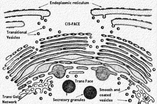
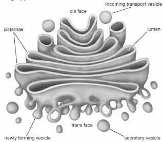
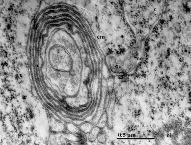
Electron microscope observations of thin sections reveal the presence of the three membranous components: (1) flattened sacs or cisternae, (2) small tubules and vesicles and (3) large vacuoles filled with an amorphous or granular substance. These membranous structures are characterized by the absence of ribosome, i.e., they are stomach membranes. These membranes structure are characterized by the absence of ribosome, i.e., they are smooth membranes.(1) The cisternae or lamellae are the most constant elements of the Golgi complex. They consist of flattened, parallel sacs pile one upon the other to form stacks.
The number of cisternae in a stack varies from species to species, and sometimes from one development stage to another. The forming or proximal face is on the outer side while the maturing or distal face is on the opposite side. It is believed that the smooth membranes of the ER bud off vesicles, which then become arranged on the forming face of the stack. It is also possible that a lamella of the ER may be converted into a Golgi lamella by loss of ribosome. On the other side is the maturing or secreting face where the lamellae bud off secretory vesicles. Thus new lamellae are formed on the forming face and mature lamellae are lost on the maturing face. Chemical analysis shows the composition of the Golgi complex membranes is intermediate between those of the ER and the plasma lemma. The membranes at the forming face of the Golgi complex are similar to the ER membranes and those of the maturing face to the plasmalemma. The membranes of the Golgi complex are in dynamic equilibrium. They are continually receiving lamellae through budding of vesicles from the smooth ER, and losing membranes through formation of secretory vesicles.
…..
Fenestration. The membranes of the cisternae may sometimes be fenestrated, i.e., may have small pores. The pores may be confined to the periphery or may occupy three-fourth or more of the cisterna. The cisternae are often reticulate structures and not simply flattened sacs. Reticulation of the cisternae may vary from one face of the stack to the other, or may also vary with different development stages.
Intercisternal elements. In a few cases dense material has been found between the cisternae of the Golgi complex, and has been called the intercisternal material. In rabbit epididymes cells the Golgi stacks are closely associated to form a continuous tube. Some stacks are also found within the tube.
(2) The small vesicles are 400-800A in diameter. They are intimately associated with the cisternae and may show continuity with them. The small vesicles arise from the cisternae by budding or pinching off.
(3) Large vacuoles. The large vacuoles are clear, and generally lie at the edge of the Golgi complex. The represent modified and expanded cisternae in which the two membranes have become widely separated, and the vacuolar space enlarged.
For more help in Electron Microscope Structure please click the button below to submit your homework assignment.